Viscoelastic Response (VisR) Imaging
Background
Viscoelastic Response (VisR) imaging, a non-ionizing and non-invasive ultrasonic imaging technology, used to estimate the viscoelastic properties of tissue. VisR is performed by exciting tissue with multiple acoustic radiation force (ARF) impulses. The resulting displacement is then captured, using standard ultrasonic motion tracking techniques, and fit to the mass-spring-damper (MSD) model. An animation of this procedure is shown in Figure 1.
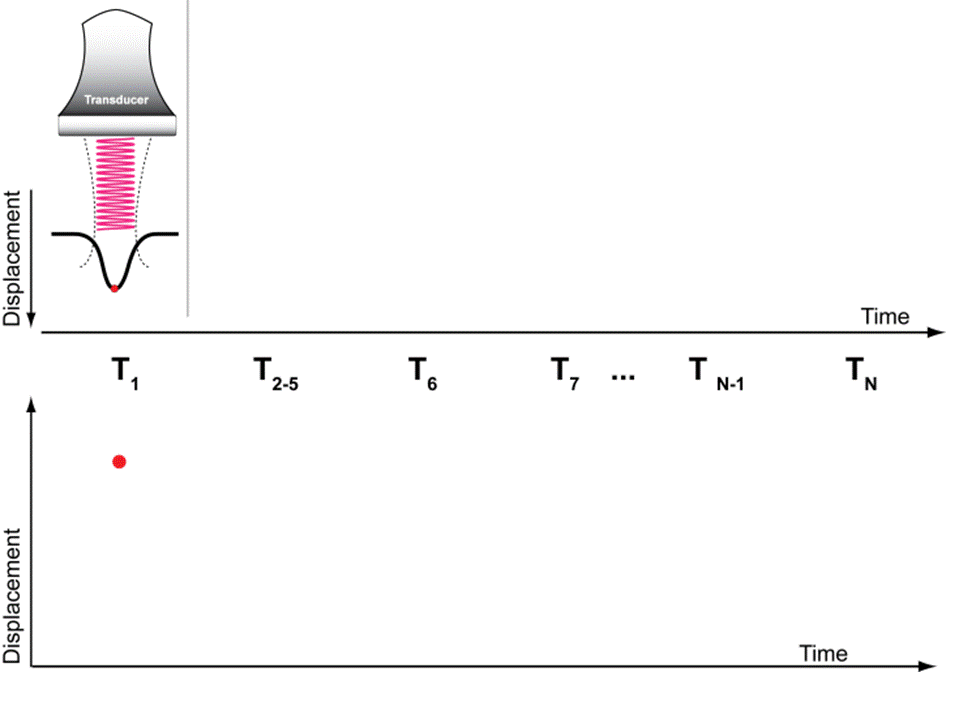
Figure 1. Animation of VisR beam sequence. Points on the plot show samples and the black line represents the MSD model fit to the data.
The displacement fit to the MSD model yields the viscoelastic parameter, τ, defined as the ratio of viscosity over elasticity. Images of τ can then be formed spanning a lateral field of view, similar to that of an ARFI image.
VisR Imaging – Finite Element Simulation
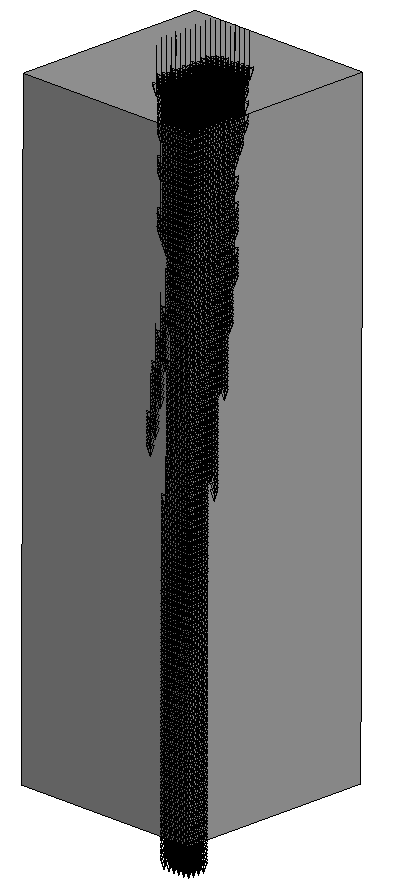
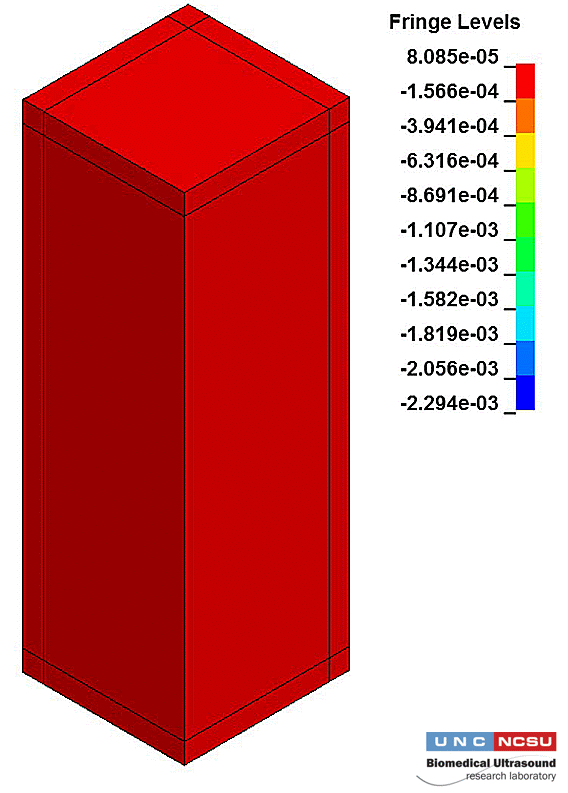
Simulation of VisR imaging have empowered rapid prototyping of VisR applications in various viscoelastic and transversely isotropic viscoelastic material models. These simulations allow for VisR to be tested in a calibrated environment with known material parameters. This calibrated environment allows us to assess fundamental limitations and sources of potential error, without the overhead of creating phantoms and implementing custom sequences on ultrasound scanner hardware. Figure 3.a shows one quarter of a finite element mesh with the ARF loading function superimposed as arrows in order to illustrate the geometry of the excitation. Panel b shows a movie of the resulting displacement from a 2-Push VisR excitation sequence using the geometry shown in a. In the movie, the color gradient ranges from low (red) to high (blue) displacement.